TORONTO – A thinner-than-normal layer of retinal nerve fibers in the eye is now linked with cognitive decline – another suggestion that extracranial physical findings could be leveraged into dementia screening tools.
The findings were seen in a cohort of 32,000 people enrolled in the U.K. Biobank– an ongoing prospective study following half a million people and collecting data on cancer, heart diseases, stroke, diabetes, arthritis, osteoporosis, eye disorders, depression, and dementia.
The correlation between retinal nerve fiber thickness and cognition was observed in the large cohort at baseline, Fang Sarah Ko, MD, said during a press briefing at the Alzheimer’s Association International Conference 2016. But after following 1,251 of these subjects for 3 years, she and her colleagues found that the correlation continued unabated.
 Courtesy National Eye Institute
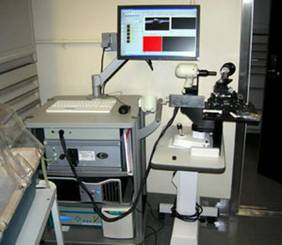
Optical coherence tomography machine used to provide an overview of the retina's structure.
“It’s amazing that we found this in such a healthy population,” Dr. Ko said during the briefing. “We wouldn’t have expected in just 3 years to see any cognitive decline in this cohort, much less measurable cognitive decline with a significant association with retinal nerve fiber layer thickness.”
Dr. Ko, an ophthalmologist in private practice in Tallahassee, Fla., said later during her main presentation of the study that the finding suggests a possible role for retinal imaging as a cognitive health screen.
“Thinner nerve fiber layer was associated with worse performance on memory, reasoning, and reaction time at baseline, and with a decline in each of these tests over time,” she said. “It may be that the nerve fiber layer could be used as a biomarker,” because it is easy to observe and measure with equipment available in most ophthalmology offices. “I would say the potential for clinical use is quite high.”
The U.K. Biobank recruits all of its subjects through the U.K. National Health Service patient registry. All undergo a standard battery of numerous tests; among them are tests of cognitive function and spectral-domain optical coherence tomography (S-DOCT) of the eye. S-DOCT is an increasingly common method of imaging the retina. It produces three-dimensional images of extremely fine resolution.
The 32,000 subjects included in the baseline cohort were all free of diabetes and ocular or neurological disease, and they had normal intraocular pressure. They undertook four tests of cognition: prospective memory, pairs matching, numeric and verbal reasoning, and reaction time. The relationship between these test results and retinal nerve fiber thickness was adjusted for age, sex, race, socioeconomic status, height, refraction, and intraocular pressure.
At baseline, the mean retinal nerve fiber layer was significantly thinner among subjects with abnormal scores on any of the cognitive tests. On the prospective memory test, the layer was an average of 53.3 micrometers for subjects who had correct first-time recall, 52.5 micrometers for those with correct second-time recall, and 51.9 micrometers for those who did not recall. The layer was also significantly thinner in subjects who had low scores on pairs matching, numeric and verbal reasoning, and reaction times.
And the relationship between test results and retinal nerve fiber thinning appeared additive, Dr. Ko said. For each test that a subject failed, the layer was about 1 micrometer thinner. In the multivariate analysis, thinner retinal nerve fiber layer was associated with worse performance on all of the tests: The layer was 0.13 micrometer thinner for each incorrect match on pairs matching; 0.14 micrometer thinner for every 2 points lower in score on numeric and verbal reasoning; and 0.14 micrometer thinner for every 100 millisecond slower reaction time.
The 3-year follow-up data confirmed that these baseline findings persisted, and predicted cognitive decline. “Again, this was true after controlling for all the variables,” Dr. Ko said. “We found that those with the thinnest layers at baseline got worse on more of the tests, compared to those who had the thickest nerve fiber layers at baseline.”
Although this is the first time retinal nerve fiber thickness has predicted cognitive decline, the association with cognition has been studied for a few years. A 2015 meta-analysis found 17 studies comparing the marker between patients with Alzheimer’s and healthy controls and 5 studies of patients with mild cognitive impairment MCI) and healthy controls (Alzheimers Dement (Amst). 2015 Apr 23;1[2]:136-43). All of these found significant retinal nerve fiber thinning in Alzheimer’s and MCI patients.
The lead author of that paper, Kelsey Thompson of the University of Edinburgh (United Kingdom), said the retinal ganglion cell axons can be seen as a sentinel marker for neurodegeneration in the brain.